Dresden researchers create liver model for improved diagnosis of non-alcoholic fatty liver disease
Non-alcoholic fatty liver disease is becoming the most common chronic liver disorder in developed countries. Histological analysis of liver tissue is the only widely accepted test for diagnosing and distinguishing different stages of the disease. However, this technique provides only two-dimensional images of the liver tissue in low resolution and overlooks potentially important 3D structural changes. Researchers at the Max Planck Institute of Molecular Cell Biology and Genetics in Dresdenand the University Hospital Carl Gustav Carus Dresden together with colleagues from the Technische Universität Dresden now generated 3D geometrical and functional models of human liver tissue for different disease stages. They reveal new critical tissue alterations, providing new insights into pathophysiology and contributing to high definition medical diagnosis.
Non-alcoholic fatty liver disease is characterized by the accumulation of fat in the liver with an insulin resistance due to causes other than alcohol intake. It includes a spectrum of liver diseases from simple steatosis (“non-progressive” and reversible) to non-alcoholic steatohepatitis, which can progress into cirrhosis, liver cancer, or liver failure, requiring eventually transplantation. In 2017, an estimated 24 percent of the worldwide population was affected by the disease, which makes it the leading cause of chronic liver disease.
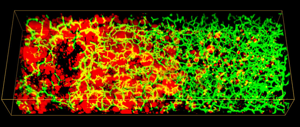
Conventional histological analysis of liver tissue is the gold standard for diagnosing disease progression, but it has several disadvantages: The low resolution 2D images of liver tissue only allow a semi-quantitative evaluation and it can be subjective, since it depends on the pathologist’s skills. Most importantly, it fails to provide 3D information on tissue structure and function, as the liver has a complex 3D tissue organization: It consists of functional units, the liver lobuli, containing two intertwined networks, the sinusoids for blood flow and the bile canaliculi for bile secretion and flux. Such an architecture makes it difficult to grasp the 3D organization and overall tissue structure from 2D histological images.


