New research shows thermal imaging techniques can predict whether a wound needs extra management, offering an early alert system to improve chronic wound care.
Nearly half a million Australians suffer from chronic wounds, which cost the nation’s health system about $3 billion each year. Untreated chronic wounds are the leading cause of limb amputation.
The Australian study shows textural analysis of thermal images of venous leg ulcers (VLUs) can detect whether a wound needs extra management as early as week two for clients receiving treatment at home.
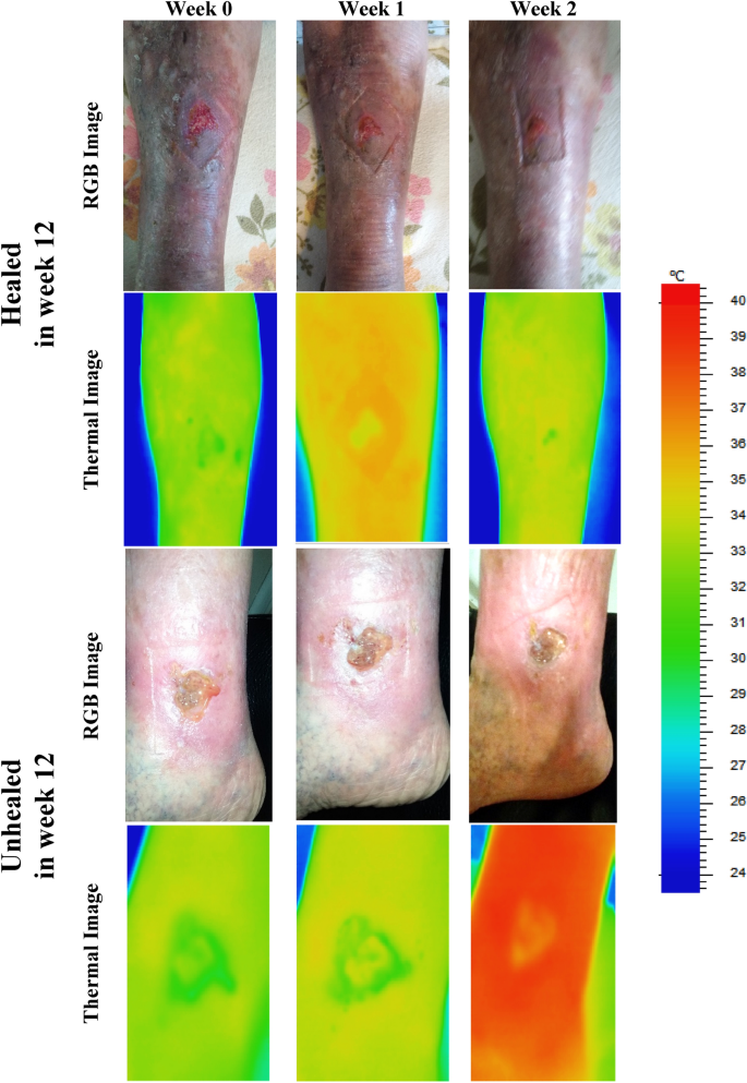
The clinical study by Bolton Clarke and RMIT University, published in the Nature journal Scientific Reports, is the first to investigate textural analysis on VLUs using thermal images that do not require physical contact with the wound.
Researchers found the method, which provides information on spatial heat distribution in a wound, could accurately predict whether VLUs would heal in 12 weeks by the second week after baseline assessment.
This is because wounds change significantly over the healing trajectory, with higher temperatures signalling potential inflammation or infection while lower temperatures can indicate a slower healing rate due to decreased oxygen in the region.
Bolton Clarke Research Institute Senior Research Fellow Dr Rajna Ogrin said the current gold standard for predicting healing of VLUs – conventional digital planimetry – requires physical contact.
“A non-contact method like thermal imaging would be ideal to use when managing wounds in the home setting to minimise physical contact and therefore reduce infection risk,” Dr Ogrin said.
After showing that traditional thermal imaging methods do not give reliable results, the research team developed a new method for the analysis and used this in the clinical trial.
The new study, which involved 60 participants with VLUs, found thermal imaging offers an improvement on the current guidance for using digital imagery or planimetry wound tracings to detect the healing wounds by week four.
“The significance of this work is that there is now a method for detecting wounds that do not heal in the normal trajectory by week two using a non-contact, quick, objective and simple method,” Ogrin said.
RMIT University Professor Dinesh Kumar said regular wound photography could not easily be used for accurate measurement of changes in wound size and other physiological parameters over time in the home care environment.
“This is because there are large variations between images due to changes in the lighting conditions, image quality and differences in camera angle across specific points in time,” he said.
“Textural analysis of thermal images is resilient to these variations and is a time-efficient and cost effective method to identify delayed healing of VLUs and improve patient outcomes.”
The research, Thermal imaging potential and limitations to predict healing of venous leg ulcers, is available here (DOI: 10.1038/s41598-021-92828-2)