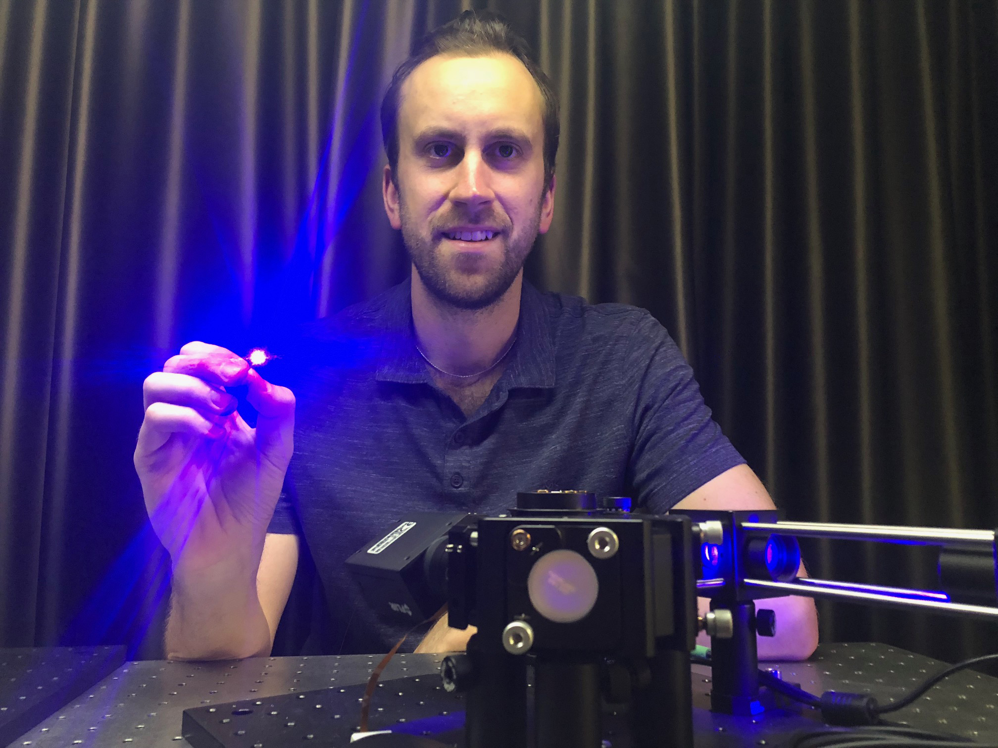
 Dr Antony Orth holding an ultra-thin microendoscope used in the study, which revealed the 3D imaging potential of the existing technology.
Researchers have shown that existing optical fibre technology could be used to produce microscopic 3D images of tissue inside the body, paving the way towards 3D optical biopsies.
Unlike normal biopsies where tissue is harvested and sent off to a lab for analysis, optical biopsies enable clinicians to examine living tissue within the body in real-time.
This minimally-invasive approach uses ultra-thin microendoscopes to peer inside the body for diagnosis or during surgery, but normally produces only two-dimensional images.
Research led by RMIT University in Melbourne, Australia, has now revealed the 3D potential of the existing microendoscope technology.
Published in Science Advances, the development is a crucial first step towards 3D optical biopsies, to improve diagnosis and precision surgery.
Lead author Dr Antony Orth said the new technique uses a light field imaging approach to produce microscopic images in stereo vision, similar to the 3D movies that you watch wearing 3D glasses.
“Stereo vision is the natural format for human vision, where we look at an object from two different viewpoints and process these in our brains to perceive depth,” said Orth, a Research Fellow in the RMIT node of the ARC Centre of Excellence for Nanoscale BioPhotonics (CNBP).
“We’ve shown it’s possible to do something similar with the thousands of tiny optical fibres in a microendoscope.
“It turns out these optical fibres naturally capture images from multiple perspectives, giving us depth perception at the microscale.
“Our approach can process all those microscopic images and combine the viewpoints to deliver a depth-rendered visualization of the tissue being examined – an image in three dimensions.”
/igwg7-meeting-overview.tmb-768v.jpg?sfvrsn=1c01de5d_1)


