Nanoscale microscopy, a tool relied on by scientists tackling tough health challenges, will be more accessible and affordable, thanks to a team of university researchers.
The University of Queensland’s Dr Martin Ploschner said super-resolution microscopy allowed users to observe breathtaking details of nanoscale structures, but for many labs, it was a costly addition to their usual microscopes.
“Standard optical microscopes can image cells and bacteria but not their nanoscale features which are blurred by a physical effect called diffraction,” Dr Ploschner said.
“Optical microscopes have evolved over the last two decades in order to bypass this diffraction limit; however, these so-called super-resolution techniques typically require expensive and elaborate instrumentation or imaging procedures.”
Dr Ploschner and his team discovered a way to bypass the usual complexities and costs of super-resolution nanoscale microscopy by using a “super-linear” fluorescent marker.
Fluorescent marker absorbs light of a certain colour and re-emits the light of a different colour.
Normally, the brightness of the emitted light is proportional to the absorbed light, but the brightness grows at a much faster rate for “super-linear” markers.
As a result, when a laser beam scans a super-linear marker, it is only the brightest, central part of the beam that causes significant glow from the fluorescent marker.
This results in sharper resolution, as the size of the emitting region is smaller than the beam itself.
“Our key discovery is that if this effect is exploited under the right imaging conditions, any standard scanning optical microscope can spontaneously image with super-resolution, at practically no extra cost,” Dr Ploschner said.
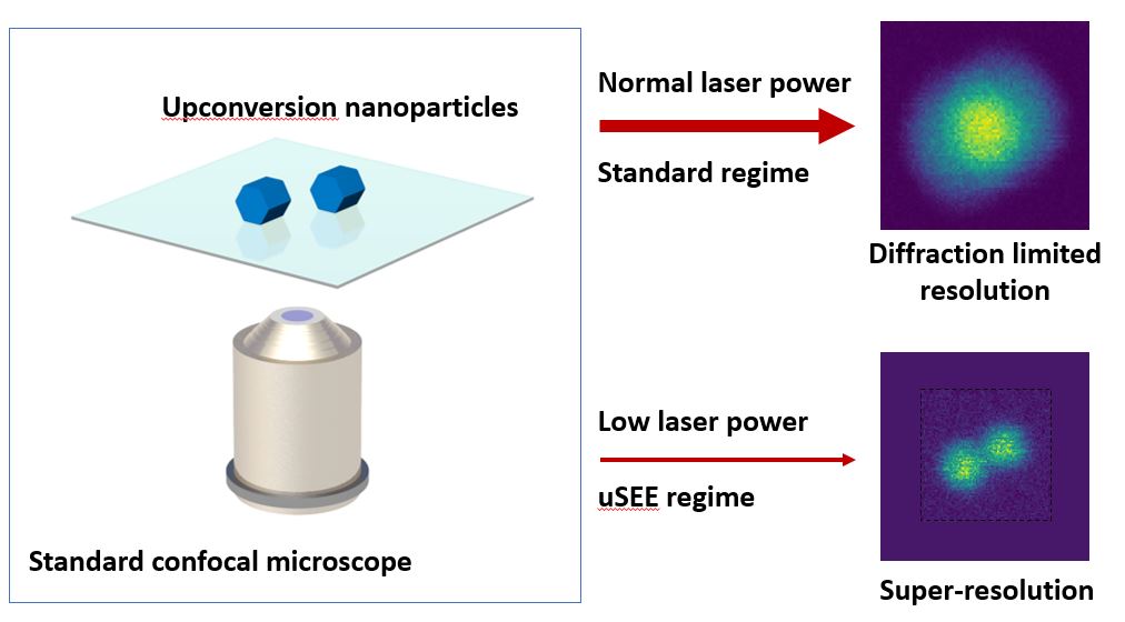 “Interestingly, our technique offers better resolution at relatively low laser power.
“This, coupled with the use of near-infrared laser, makes the technique appealing for imaging of biological samples,” he said.
The team believes the approach had the potential to open new avenues in the research of super-liner emitters, combining them with other imaging processes to improve their performance.
Researchers from Macquarie University and ARC Centre for Excellence for Nanoscale Biophotonics worked on 3D sub-diffraction imaging in a conventional confocal configuration by exploiting super-linear emitters, published in Nature Communications (doi.org/10.1038/s41467-019-11603-0).
This work was supported by the Australian Research Council funding through the Centre for Excellence for Nanoscale Biophotonics.


