
Scientists have developed a new graphene-based brain monitoring technology that could help doctors better understand what happens in the brain during a stroke and identify areas at risk of further damage.
The research, published in Brain, brought together experts in neuroscience, stroke research, engineering and advanced materials from the University of Manchester, the Institute of Microelectronics of Barcelona (IMB-CNM, CSIC) and the Catalan Institute of Nanoscience and Nanotechnology (ICN2) in Barcelona, together with industry partner Multi Channel Systems in Germany.
When a person has an ischaemic stroke, a blockage cuts off blood supply to part of the brain. However, the damage does not stop immediately. Waves of disrupted electrical activity, known as cortical spreading depolarizations, travel through the injured brain and can cause additional harm. Until now, it has been difficult to measure these signals accurately because existing technologies poorly capture the very slow electrical changes involved.
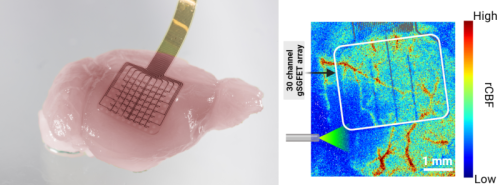
Using highly sensitive graphene-based sensors placed on the brain, researchers were able to record these electrical waves in unprecedented detail. They discovered that the shape of the electrical signals revealed important information about the health of the surrounding brain tissue. The signals could distinguish between tissue that was relatively healthy, tissue that was at risk, and tissue that was already severely compromised. These tests were performed in mice.
The researchers also found that these signals could predict how blood flow would respond to the electrical wave. In healthier tissue, blood flow increased to help the brain recover from the event, whereas in vulnerable tissue blood flow could instead further decrease, worsening the injury.
Importantly, the researchers showed that these harmful responses could be altered using a low dose of ketamine, a drug already used in medicine. Ketamine reduced the duration of the damaging electrical waves, shifted the associated blood flow response from potentially harmful vasoconstriction towards vasodilation, and reduced the overall area of brain damage. These findings reveal one potential mechanism that may contribute to the neuroprotective effects of ketamine following stroke.
The findings suggest that graphene-based brain monitoring could one day provide doctors with a real-time window into the condition of brain tissue during a stroke. This could help identify patients at greatest risk of further injury and support the development of new treatments aimed at protecting the brain and improving recovery.
Dr Rob Wykes, Senior Lecturer at The University of Manchester, said: “More broadly, the study demonstrates how cutting-edge technologies can not only improve the way brain activity is measured but also reveal important new insights into how brain injuries develop and how they might be treated.”
The study’s first author was Dr Samuel Flaherty from The University of Manchester. Corresponding authors were Dr Anton Guimerà-Brunet from the Institute of Microelectronics of Barcelona (IMB-CNM, CSIC) and Dr Rob Wykes from the University of Manchester and UCL Queen Square Institute of Neurology.
“Recording these ultraslow signals remains highly challenging with current technologies. We have been developing our graphene-based technology for years, with our first results published in 2018, and the challenge has been to standardise it and make it mature for use in relevant preclinical research,” states Dr Guimerà-Brunet, from IMB-CNM.
A consolidated collaboration
This advance marks a new milestone in the long-standing collaboration between IMB-CNM, the University of Manchester and ICN2, which began with the development of graphene microtransistor technology for recording ultraslow brain signals, first published in Nature Materials in 2018.
Since then, the teams have continued to develop graphene-based technologies for neural interfaces, advancing both the recording of brain signals across a wide range of frequencies and devices capable of combining neural recording and stimulation, with studies published in Nature Nanotechnology and, more recently, in Nature Communications. These technologies are developed and validated in collaboration with specialised research groups to explore their application beyond the laboratory.
This research was published in: Brain
Full title of the paper: “Monitoring ischaemic tissue vulnerability through perfusion-dependent signatures of spreading depolarizations”
DOI: 10.1093/brain/awag305
URL: https://academic.oup.com/brain/advance-article/doi/10.1093/brain/awag305/8788779



-fao-lebanon--ralph-azar--fao-delegation-official-travel-beirut-lebanon_0225s.jpg?sfvrsn=1849021a_6)