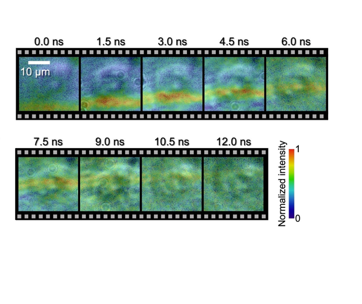
Images of an underwater shock wave moving through a HeLa cell. Using this new technology, the researchers could see the difference between how the shock wave moved inside and outside of a cell submerged in water. They noted that the results suggested that the cell structure shifts with the visualized wavefront position (shown in the red/ orange line in the image). © 2023 Saiki et al.
A microscopic shock wave has been photographed passing through a single biological cell, thanks to a new photography technique. Nanosecond photography uses ultrafast electronic cameras to take images at the speed of a billionth of a second. However, image quality and exposure time are typically limited. Now, a team led by researchers at the University of Tokyo has achieved superfine images taken over multiple timescales at high speed using a system they named spectrum circuit. Spectrum circuit bridges the gap between optical imaging and conventional electronic cameras, enabling photography at ultrafast speeds with less blur and more accuracy. This technology has potential applications for science, medicine and industry.
You’re waiting with your camera for just the right moment. Suddenly, your subject speeds by and you’ve barely clicked the shutter. Missed it. Timing can be everything in photography and capturing images at high speed poses a particular challenge. But thanks to advances in camera technology, these days we can see the world like never before. Whether it’s the sweat on a racing cyclist’s brow, the focus in the eyes of a swooping falcon or, with this latest improvement in nanosecond photography, the movement of a shock wave passing through a microscopic single cell at high speed.
“For the first time in history, as far as we know, we have directly observed the interaction between a biological cell and a shock wave, and experimentally demonstrated that the velocity of the shock wave propagating inside the cell is faster than the outside of the cell,” explained Takao Saiki, a doctoral student from the Department of Precision Engineering at the University of Tokyo. “Furthermore, our approach has enabled us to demonstrate high-speed photography across a wide time range, which includes picosecond (one-trillionth of a second), nanosecond (one-billionth of a second) and millisecond (one-thousandth of a second) timescales.”
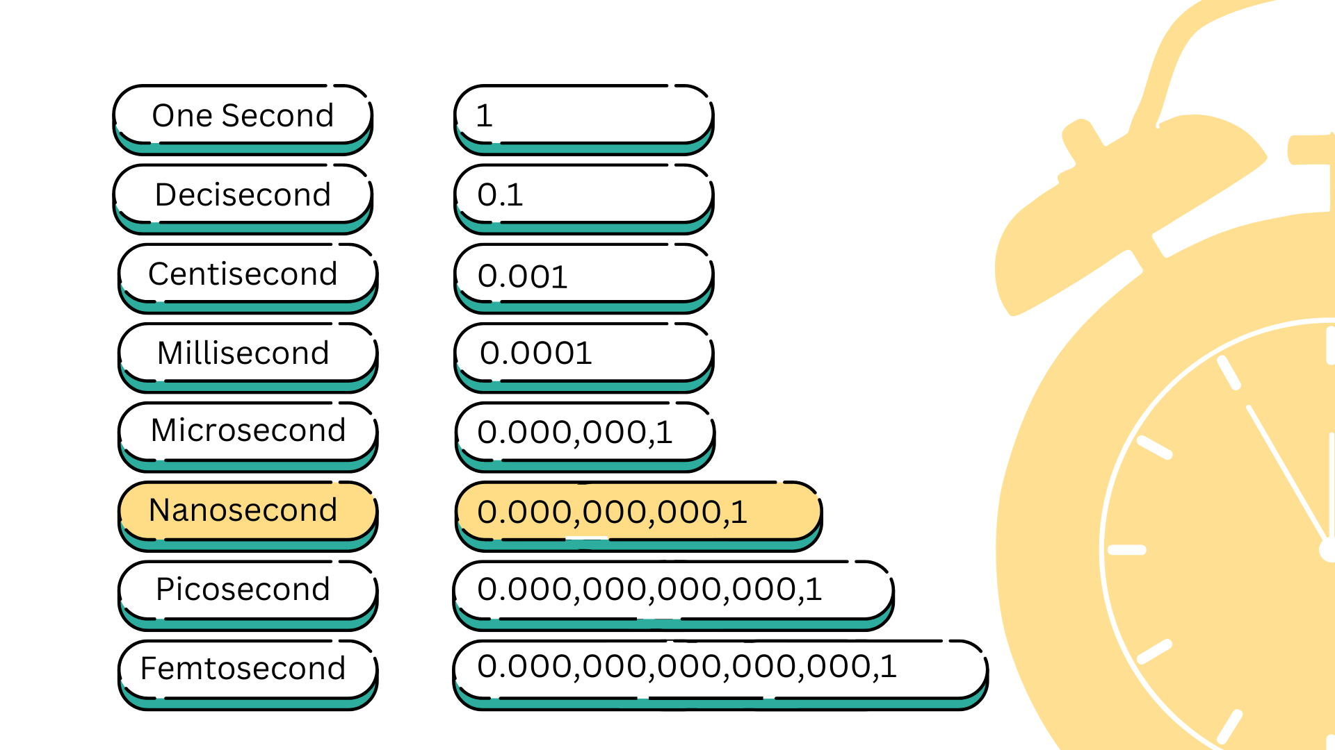
Less than a second. Picoseconds are the typical rate used in ultrafast optical imaging, while high-speed electronic cameras can take images at the rates of millisecond and microsecond. The research team’s spectrum circuit system bridges the gap between these technologies, enabling us to see what happens in between these time frames. © 2023 Nicola Burghall
Capturing clear images of cells without affecting their structure or causing damage is very challenging. To safely take the images, the researchers developed a precision optical circuit, a circuit that uses light instead of electricity, which they named spectrum circuit. With spectrum circuit they created nondamaging laser pulses, which they set to emit at different timings. By combining this technology with an existing single-shot optical imaging technique called sequentially timed all-optical mapping photography, or STAMP, they were able to take series of images with higher definition and less blur than previously available.
The team used the same technology to look at the effects of laser ablation on glass. Laser ablation is useful for precisely removing solid material from a surface and is used in both industry and medicine. The researchers focused an ultrashort laser pulse just 35 femtoseconds long (one femtosecond is equal to one-quadrillionth of a second) onto a glass plate. Using the spectrum circuit, they observed the impact of the laser, the resulting shock waves, and the effect it had on the glass over picoseconds, nanoseconds and milliseconds.
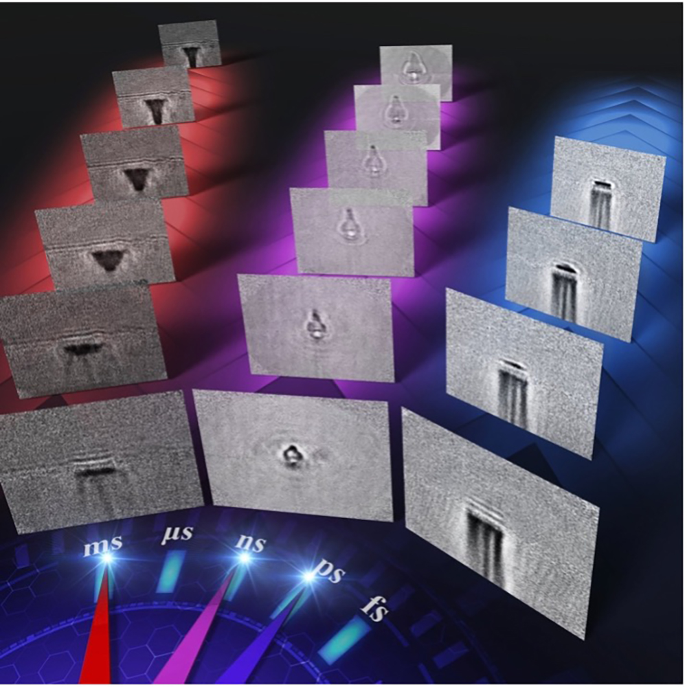
Images of laser ablation taken using the ultrawide time range, high-speed camera. Applying this new imaging technology, the researchers could see the propagating shock wave and plasma and the progress of laser processing over multi-timescales (about 10–100 picoseconds, about 1–10 nanoseconds, and about 1–100 milliseconds). © 2023 Saiki et al.
“We could see the interplay between different physical processes taking place over time, and how they took shape,” said Keiichi Nakagawa, an associate professor from the Department of Bioengineering and the Department of Precision Engineering at the University of Tokyo. “Our technology provides opportunities to reveal useful but unknown high-speed phenomena by enabling us to observe and analyze such ultrafast processes.
“Next, we are planning to use our imaging technique to visualize how cells interact with acoustic waves, like those used in ultrasound and shock wave therapy. By doing this, we aim to understand the primary physical processes that activate subsequent therapeutic effects in the human body.” The team also wants to use spectrum circuit to improve laser processing techniques, by identifying the physical parameters that would enable faster, more precise, more consistent and cost-effective manufacturing.
“We have always been fascinated by the power of visualization to understand complex phenomena. The chance to uncover and show parts of the world that were hidden before really drew us to this field,” said Nakagawa. “We expect to make broad contributions in various fields, from biomedicine to manufacturing, materials, the environment and energy.”


