Changes in the eye could signal Alzheimer’s disease, according to new research showing that physical changes in cells of the retina can occur at the same time as brain changes found in the early stages of Alzheimer’s disease.
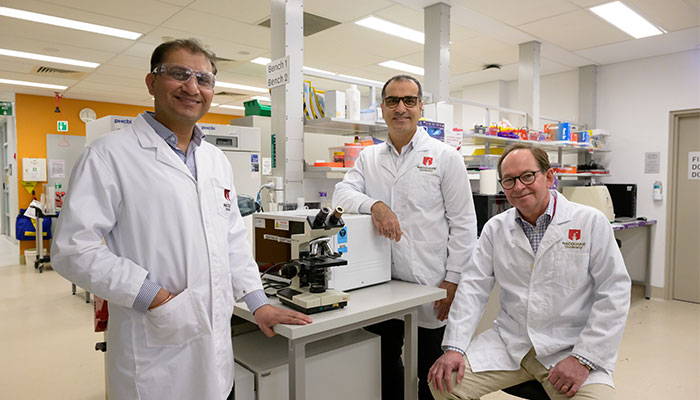
Advancement: Researchers Dr Vivek Gupta, Dr Mehdi Mirzaei and Dr Stuart Graham (pictured left to right) developed a world-first protein composition map of human retinas which contributed to the findings of a major study linking changes in the retina to Alzheimer’s disease.
A major international study into shared molecular markers and pathways by brain and eye in Alzheimer’s disease, has used the world’s first ‘proteome map’ of both eyes and brains concurrently affected by Alzheimer’s disease, developed by Macquarie University researchers.
“Researchers have recognised in recent years that there could be changes in the retina quite early in the disease process, but trying to identify these in the eyes of live patients has proved tricky,” says Professor Stuart Graham, head of Ophthalmology and Visual Science at Macquarie University.
Professor Graham says that research at Macquarie is investigating how two proteins, called– beta-amyloid and tau, which are known to build up in brains of Alzheimer’s patients, can also be found in cellular tissue of the eye, often long before symptoms are apparent.
This research could potentially contribute to the future development of an imaging technique with the potential to detect Alzheimer’s disease with a non-invasive eye test.
“The accumulation of beta-amyloid and tau in the central nervous system might be a key initiating factor in the development of Alzheimer’s disease,” says Associate Professor Vivek Gupta, a visual neurobiologist who leads Macquarie’s Vision Neurodegeneration research group.
He says while the specific cause of Alzheimer’s disease remains unclear, “recent studies by our group and others have reported similar pathological processes and alterations in the retina.”
International study
Together with proteomics expert Associate Professor Mehdi Mirzaei from Macquarie University’s Vision Science team, Professor Graham and Associate Professor Vivek Gupta have contributed to an important new international study which analysed donor brain and retina tissue from 86 people.
Hope: Macquarie researchers says findings of the seven-year international study brings us closer to developing future treatments for Alzheimer’s disease.
The group developed a holistic proteome map of human retinas, and of the brains of people who had Alzheimer’s disease, showing protein changes at the molecular, cellular and structural levels of both eye and brain, and associated cell death and inflammation.
Donors included people with normal brain function, some with mild cognitive impairment, and others who were diagnosed with Alzheimer’s disease; and 39 people had donated both retina and brain tissue, so researchers could directly compare protein levels in each.
The research spanned seven years and involved scientists from 18 different institutions in Australia, Italy and the US, headed by neuroscientist Maya Koronyo-Hamaoui, from Cedars Sinai Medical Center in California.
Disease biomarkers
The international study found that people who had Alzheimer’s disease, showed nine times the amount of beta-amyloid protein in their retina compared to people who didn’t show any signs of cognitive impairment during their lifetime.
This protein is a specific marker of Alzheimer’s disease; and the study also found that these markers occurred at around five times the rate in people who had not been diagnosed with Alzheimer’s disease during their lifetime, but had shown signs of mild cognitive impairment.
This underscores the immense potential of further studies on the proteome of the eye and brain.
The researchers were also able to track patterns in the location of these markers within the retina, with higher levels found in the tissues of the retina’s inner layer.
“We don’t have a device in the clinic to identify these changes in a living eye yet; but if we can label these proteins, then develop an imaging device that can spot change at the earliest stages, we may have a way to clinically diagnose diseases such as Alzheimer’s,” says Professor Graham.
Common pathways
Professor Graham says that a range of degenerative diseases (including Alzheimer’s) share common cell deterioration pathways, and identifying patterns in the types and locations of abnormal proteins in retinal cells may also help diagnose such diseases as glaucoma.
“We can readily diagnose advanced glaucoma now by imaging the back of the eye, but we also know there are changes occurring at the molecular or cellular level, long before we can see those structural changes when nerve fibres start to drop out,” he says.
The sooner a diagnosis is made in degenerative disease it means treatments can be targeted at stopping the damage at an early stage.
One of the keys to our ability to identify the progress of different degenerative diseases through changes in the eye, is likely to be via understanding the ‘proteomics’ or the protein make-up of cells in the retina.
Protein Bars
The development of a proteome map involved a detailed analysis of brain and eye tissue at molecular level, and drew on Macquarie University’s deep proteomics expertise.
“Proteomics is the study of the entire set of proteins present in a particular tissue or biological sample level,” says Associate Professor Mirzaei.
He says that quantitative proteomics has become a powerful tool in understanding, diagnosing and treating disease. It can help find markers to identify and predict diseases and mark disease progression. It can reveal potential new medicine targets, be used to create personalised treatments and even show the effectiveness of disease treatments.
“We’ve been applying these techniques in vision neuroscience for the past few years, in collaboration with Stuart Graham’s group, and with a range of other internal and external partners,” Associate Professor Mirzaei says.
“Initially we focused on identifying the molecular mechanisms that were apparent in glaucoma, which paved the way for our ground-breaking research into deciphering the shared molecular markers and pathways in Alzheimer’s disease.”
Associate Professor Mirzaei says their work was meticulously compared against other crucial Alzheimer’s brain datasets available online, and also unearthed a remarkable similarity between human tissues and animal models of the disease.
“This underscores the immense potential of further studies on the proteome of the eye and brain in various animal models of Alzheimer’s disease, bringing us closer to developing future treatments,” Associate Professor Mirzaei says.
Professor Stuart Graham is Head of Ophthalmology and Visual Science at Macquarie University.
Dr Mehdi Mirzaei is an Honorary Associate Professor at the Macquarie Medical School.
Dr Vivek Gupta is an Associate Professor at the Macquarie Medical School.


