
What the research is about
Ulcerative colitis causes symptoms such as abdominal pain, diarrhea, and bloody stools. In this disease, the mucosal lining of the colon-the main part of the large intestine-becomes inflamed and develops open sores called ulcers. When the inflammation continues, the intestinal lining goes through repeated cycles of injury and repair.
It has long been known that Paneth cells, which are normally found mainly in the small intestine, can also appear in the colon of patients with ulcerative colitis. This change is called Paneth cell metaplasia. It has been reported since at least the 1960s and was long regarded simply as a tissue change caused by chronic inflammation. However, researchers did not know why these cells appeared in the colon or what role they played in the injured intestine.
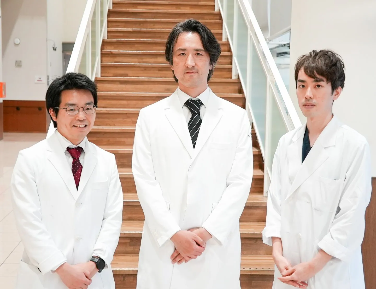
To understand why these cells appear and what they do, a research team led by Assistant Professor Go Ito and graduate student Tomohiro Muto, now a Project Researcher at Institute of Science Tokyo (Science Tokyo), examined tissue samples from patients with ulcerative colitis. In addition to tissue analysis, the researchers combined several experimental approaches, including mouse models and organoids-miniature intestine-like structures grown from patients’ colon cells.
Why this matters
Analysis of colon tissue showed that Paneth cell metaplasia was more common in patients who had lived with ulcerative colitis for more than ten years. It was also found more frequently in areas with active inflammation. However, these Paneth-like cells were very rare in patient tissues, making them difficult to study in detail. The team therefore used a recently developed technology, called spatial transcriptomics. This technology enables researchers to examine gene activity while preserving the original architecture of the tissue. It allowed the team to compare normal Paneth cells from the small intestine with the Paneth-like cells found in the colon.
Paneth cells release substances called antimicrobial peptides, which help control gut bacteria and maintain a healthy balance in the intestinal microbiome. The study showed that the Paneth-like cells in the colon produced a different combination of antimicrobial peptides compared with normal Paneth cells in the small intestine. This suggested that the cells had developed characteristics adapted to the inflamed environment. To understand this adaptive response of the cells, the researchers focused particularly on IL-22, a signaling protein released by immune cells during inflammation, and REG3A, an antimicrobial peptide whose production is stimulated by IL-22.
The team then combined human tissue analysis, organoids grown from human and mouse colon cells, and mouse models. Human tissue samples revealed the characteristics of the cells. The organoids showed how the cells changed and how REG3A acted, while the mouse models allowed the researchers to examine the role of REG3A in wound repair.
The results showed that IL-22 acts as a trigger for Paneth cell metaplasia in the colon. The resulting Paneth-like cells release REG3A, which promotes the growth of cells lining the colon and helps wounds close. These findings suggest that Paneth cell metaplasia, long considered simply a tissue change caused by prolonged inflammation, may not simply be a consequence of chronic inflammation. Instead, it may be an adaptive response that helps the injured intestine repair itself.
What’s next
Harnessing the effects of REG3A could lead to new treatments that not only suppress inflammation but also actively promote the repair of damaged intestinal tissue. Paneth cell metaplasia may also serve as a pathological marker of accumulated chronic inflammation that cannot always be detected through endoscopy alone. In the future, it could provide useful information for long-term disease management and the assessment of colorectal cancer risk.
Comment from the researcher
Whenever I saw the term ‘Paneth cell metaplasia’ in the pathology reports of patients with ulcerative colitis, I wondered, ‘Why are cells appearing where they do not normally belong?’ I felt that even changes that look abnormal must carry some biological meaning, and that question motivated me to continue this research.
This discovery is a starting point for exploring a fundamental question in biology: How far can cells change their form and function in response to their environment? We hope this work will also deepen our understanding of the roles played by different forms of metaplasia that arise during chronic inflammation.
(Go Ito, Assistant Professor, Department of Gastroenterology and Hepatology/ The Center for Personalized Medicine for Healthy Aging, Institute of Science Tokyo)
Dive deeper
Related research by Assistant Professor Go Ito
Explore more research in Science for All
Science for All showcases cutting-edge research at Science Tokyo and highlights the ideas, people, and possibilities shaping the future.