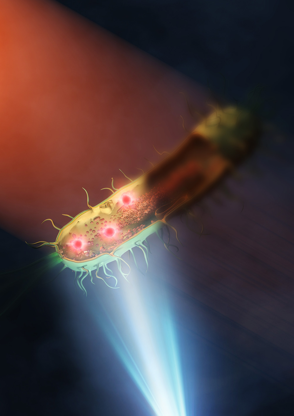
Illustration of new mid-infrared technique. This illustration represents a bacteria being illuminated with mid-infrared in the top left, while visible light from a microscope underneath is used to help capture the image. © 2024 Ideguchi et al./ Nature Photonics
A team at the University of Tokyo have constructed an improved mid-infrared microscope, enabling them to see the structures inside living bacteria at the nanometer scale. Mid-infrared microscopy is typically limited by its low resolution, especially when compared to other microscopy techniques. This latest development produced images at 120 nanometers, which the researchers say is a thirtyfold improvement on the resolution of typical mid-infrared microscopes. Being able to view samples more clearly at this smaller scale can aid multiple fields of research, including into infectious diseases, and opens the way for developing even more accurate mid-infrared-based imaging in the future.
The microscopic realm is where viruses, proteins and molecules dwell. Thanks to modern microscopes, we can venture down to see the inner workings of our very own cells. But even these impressive tools have limitations. For example, super-resolution fluorescent microscopes require specimens to be labeled with fluorescence. This can sometimes be toxic to samples and extended light exposure while viewing can bleach samples, meaning they are no longer useful. Electron microscopes can also provide very impressive details, but samples must be placed in a vacuum, so live samples cannot be studied.
By comparison, mid-infrared microscopy can provide both chemical and structural information about live cells, without needing to color or damage them. However, its use has been limited in biological research because of its comparatively low resolution capability. While super-resolution fluorescent microscopy can narrow down images to tens of nanometers (1 nanometer being one-millionth of a millimeter), mid-infrared microscopy can typically only achieve around 3 microns (1 micron being one-thousandth of a millimeter).
However, in a new breakthrough, researchers at the University of Tokyo have attained a higher resolution of mid-infrared microscopy than ever before. “We achieved a spatial resolution of 120 nanometers, that is, 0.12 microns. This amazing resolution is roughly 30 times better than that of conventional mid-infrared microscopy,” explained Professor Takuro Ideguchi from the Institute for Photon Science and Technology at the University of Tokyo.
The team used a “synthetic aperture,” a technique combining several images taken from different illuminated angles to create a clearer overall picture. Typically, a sample is sandwiched between two lenses. The lenses, however, inadvertently absorb some of the mid-infrared light. They solved this issue by placing a sample, bacteria (E. coli and Rhodococcus jostii RHA1 were used), on a silicon plate which reflected visible light and transmitted infrared light. This allowed the researchers to use a single lens, enabling them to better illuminate the sample with the mid-infrared light and get a more detailed image.
“We were surprised at how clearly we could observe the intracellular structures of bacteria. The high spatial resolution of our microscope could allow us to study, for example, antimicrobial resistance, which is a worldwide issue,” said Ideguchi. “We believe we can continue to improve the technique in various directions. If we use a better lens and a shorter wavelength of visible light, the spatial resolution could even be below 100 nanometers. With superior clarity, we would like to study various cell samples to tackle fundamental and applied biomedical problems.”


/igwg7-meeting-overview.tmb-768v.jpg?sfvrsn=1c01de5d_1)
