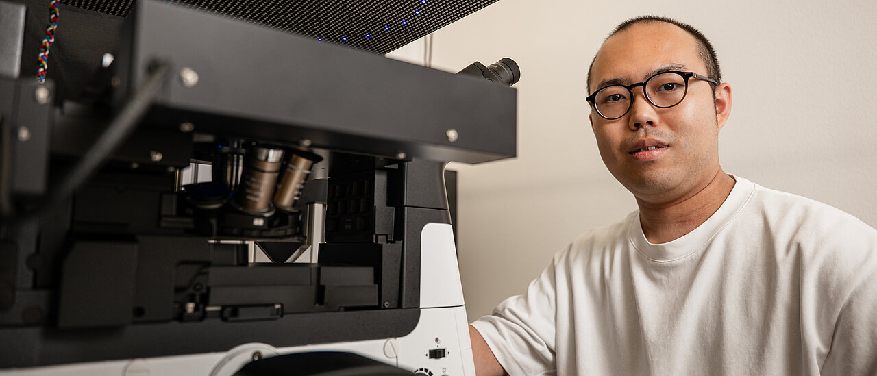
Shoma Kataoka’s goal is to accelerate the analysis of large volumes of samples, such as those involved in drug discovery. He will receive close to 220,000 euros in funding for his research in Würzburg.
Discovering new drugs in medicine requires analyzing samples exposed to different treatments – huge quantities of them. To discover just one active substance, this number can run into the tens of thousands. In one widely used approach, bacteria, cells or tissues are placed on plates containing nearly hundred or more than thousand wells, each containing a different substance, and examined. Scientists refer to this process as ‘phenotypic screening’.
During this process, researchers use microscopes to observe how the samples react, record these reactions in images, and ultimately analyze them, now often with AI methods that can detect subtle changes between wells that the human eye cannot see. However, to capture such details, conventional microscopes need high magnification objectives, which in turn restrict the size of the imaged area to only a small region within a well. As a consequence, the microscope needs to move the plate in order to scan multiple regions within each well and from one well to the next to obtain sufficient information from all samples. Due to the sheer number of samples involved, this scanning process is very time-consuming and can also generate artifacts that fool AI methods, which can become a bottleneck in drug discovery and other large-scale biological experiments.
To address this issue, Würzburg-based scientist Shoma Kataoka is developing a new microscopy system that dispenses of scanning but is still able to capture detailed images. His research project, ‘PtychoScreening – Fourier Ptychography Phenotypic Screening’, has received close to 220,000 euros in funding from the European Commission’s Marie Skłodowska-Curie Actions (MSCA). Kataoka works at the Chair of Machine Biophotonics at the University of Würzburg (JMU) as part of Professor Christophe Zimmer’s team.
High-throughput microscopy is set to become over 1,000 times faster
Kataoka’s project goal is ambitious: the microscopy platform he develops aims to image an entire well plate in less than a minute without moving the plate, while still allowing to reveal structures smaller than half a micrometre – roughly the width of a bacterial cell. The platform could be applied to bacteria, cells or tissue equally well.
“Capturing many thousands of images by scanning a plate under the microscope is time-consuming and leads to unwanted variations unrelated to the biology”, explains the researcher. “This compromises the comparability and reliability of the data and limits the rate of drug discovery. We expect that our new platform will enable us to speed up the existing AI-based phenotypic screening technique by more than a thousandfold.”
The platform is based on a technique known as Fourier-ptychographic microscopy. In this computer-assisted method, several low resolution images obtained from different illumination angles are mathematically combined to form a high-resolution composite image. This makes it possible to visualise a large area of the sample under investigation while also capturing the fine structures of bacteria or cells. However, the technique is not yet sufficient for the rapid analysis of entire well plates. Kataoka therefore intends to further develop it along three directions:
- Firstly, the samples are to be imaged in the future with a slightly adjusted focus. This will allow a specially designed software to reconstruct finer details from the resulting images than the pixel size of the camera would suggest at first glance.
- Secondly, a new computational algorithm will be designed to compensate for distortions typically caused by viewing through the walls of the sample chambers and the curved surface of the liquid within them. To achieve this, the software will take the exact shape and arrangement of the well plate into account.
- Thirdly, the number of images required is to be reduced. Kataoka intends to use a technique known as compressed sensing to achieve this. This method exploits the fact that images of biological samples contain highly redundant information, which enables reconstructing them from fewer measurements than typically believed necessary. This should enable a high-quality overall image to be obtained from significantly fewer images.
From mechanical engineering to biological imaging:
Kataoka completed his PhD in Engineering at the University of Osaka in 2025. He had previously obtained his Bachelor’s and Master’s degrees in Mechanical Engineering at the same institution. From 2023 to 2025, he was also a research fellow of the Japan Society for the Promotion of Science.
“The Marie Skłodowska-Curie grant gives me the opportunity to conduct independent research in an international environment,” he says. “I can work at the interface between imaging and biological applications, developing new skills through collaboration with other researchers. For me, this is an important step towards an independent scientific career in computer-aided imaging.”
When the grant begins in mid-August 2026, he intends to start by evaluating Fourier-ptychographic microscopy on bacteria and other biological samples and identifying the practical challenges that still need to be resolved before the system can be applied for phenotypic drug discovery.