
Scientists studying the neurology of tame foxes identified changes in a part of the brain that has previously been associated with aggression, according to a new paper that compared the brains of domesticated, aggressive and wild foxes.
The changes occurred in an area at the base of the brain called ventromedial hypothalamus, which is known to drive aggression, control decisions related to energy use and is important for maternal instincts and social behaviors in mice.
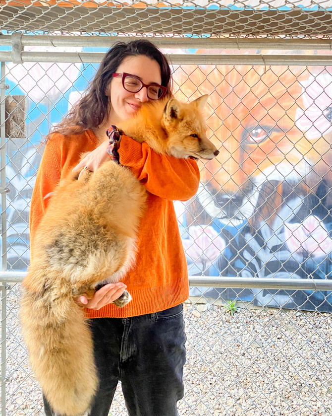
Eirene Markenscoff-Papadimitriou with a tame silver fox.
The study, published Nov. 2 in the journal Current Biology, takes advantage of a classic experiment that started in Siberia in the late 1950s where Russian geneticists noticed some foxes on silver fox farms were friendlier toward human caretakers while others were more aggressive.
They then selectively bred friendlier foxes with each other and after only six generations of this selection, they had litters of puppies that interacted with humans. The cuddlier foxes also had physical features characteristic of animal domestication, including floppy ears and shorter snouts.
“We wanted to understand, what are the neurological underpinnings of these changes in behaviors that occurred very rapidly with this selection,” said Eirene Markenscoff-Papadimitriou, assistant professor of molecular biology and genetics in the College of Agriculture and Life Sciences (CALS), a Rita Allen Foundation Scholar and the paper’s senior author.
The research has implications for how environmental pressures of all kinds can alter brain structure on short timescales, Markenscoff-Papadimitriou said.
In the study, the authors decided to focus on the ventromedial hypothalamus because previousresearch had already identified different cell types there that control aggression in mice.
Working with Russian fox colony scientists, the study’s authors received a shipment of nine silver fox brains: three from foxes bred to be tame, three from foxes similarly bred for aggressive traits and three wild foxes. The brains were preserved and very thinly sliced. In the ventromedial hypothalamus, a fluorescent RNA scope allowed them identify markers for different cell types there.
Previously, other scientists had employed a technique called optogenetics in experiments with mice where light was used to activate specific neurons that express a receptor protein called estrogen receptor alpha in the ventromedial hypothalamus.
“If you activate them, the mouse will immediately attack another mouse,” Markenscoff-Papadimitriou said. “We hypothesized that there would be differences in the abundance of this particular cell type, and we did find that the tame fox hypothalamus had fewer of these estrogen receptor expressing neurons, which are known to be part of the aggression circuit,” she said.
Now that the paper’s authors observed this difference, next steps will involve trying to understand why domestication leads to these changes in the ventromedial hypothalamus, a brain area has not been well studied before this work.
Study co-author Anna Kukekova, an associate professor of genomic biology and animal sciences at the University of Illinois-Urbana-Champaign, may explore how mutations in different parts of the fox genome affect the development of these particular neurons.
Jared Klein ’25, a former research technician in Markenscoff-Papadimitriou’s lab, is the paper’s first author. Co-authors include Andrew Miller, professor of population medicine and diagnostic sciences in the College of Veterinary Medicine; and Anastasiya Kharlamova, Anastasiya Vladirimova and Darya Shepeleva at the Institute of Cytology and Genetics of the Russian Academy of Sciences.
The study was funded by Cornell’s Center for Vertebrate Genomics.