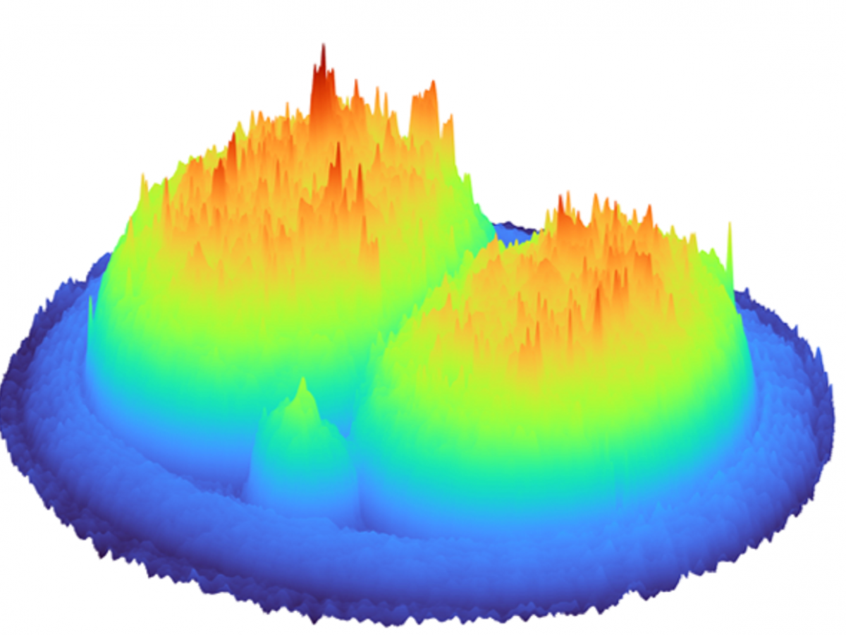
A 3D hologram of a 2-cell embryo generated by the digital holographic microscope.
In a world-first, 3D holographic images of an embryo have been developed as part of a collaborative research project between the University of Adelaide and University of St Andrews. The images are created using miniscule amounts of light in a fraction of a second.
The team, led by Dr Kylie Dunning, Hospital Research Foundation fellow from the University of Adelaide’s Robinson Research Institute, and Professor Kishan Dholakia from the University of Adelaide and the University of St Andrews, developed an approach to create 3D holographic images of the pre-clinical model of an embryo at various stages of development.
“For couples wishing to conceive, the quality, or developmental potential, of an embryo is critical as it dictates the success of their pregnancy and ultimately, the birth of their child,” said Dr Dunning.
“In vitro fertilisation (IVF) clinics routinely assess embryo quality by visual inspection to check if an embryo is developing in a time-appropriate manner or by an invasive biopsy to determine DNA content of the biopsied sample.
“However, these approaches have failed to improve the success rate of IVF which has remained stagnant for more than a decade.”
A non-invasive approach without biopsy to help pick the most appropriate embryo is a highly beneficial tool for the 21st century embryologist: light can fulfil this need.
3D holographic images are a non-invasive approach which provides insights into the embryo by identifying detailed features. This may augment conventional visual assessment for embryo quality in an IVF clinic, allowing an embryologist to make an informed decision on the selection of best quality embryos.
“Optical technologies hold immense promise to unravel the metabolism and health of the embryo. This gentle, non-invasive approach could lead to improved IVF success,” said Dr Dunning.
“Optical technologies hold immense promise to unravel the metabolism and health of the embryo. This gentle, non-invasive approach could lead to improved IVF success.”Dr Kylie Dunning, University of Adelaide’s Robinson Research Institute.
Data from 2020 show that the success rates of IVF range from a live birth rate of 38.9 per cent per embryo transfer for patients under 34 years, to a live birth rate of 5.6 per cent per embryo transfer for patients over 43 years. In 2018 it was estimated that eight million babies had been born through IVF since the world’s first in 1978.
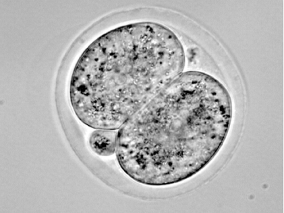
A 2D brightfield image of a 2-cell embryo. This is what an embryologist currently sees using a microscope.
“This technology uses miniscule amounts of light – less than that from your smartphone – to allow rapid visualisation of the embryo in a fraction of a second,” said Professor Dholakia.
“It’s a prime example of interdisciplinary success for our new Centre of Light for Life at the University of Adelaide, and of collaborative international work with my group at the University of St Andrews, Scotland.”
The team aims to have the technology, which is being developed through research using a preclinical model, available in five years.
This cutting-edge development would not have been possible without the support of funding from the UK and EU, and the Australian Research Council (ARC), National Health and Medical Research Council and the Hospital Research Foundation in Australia.
Primary authors on the study are George Dwapanyin, postdoctoral researcher at the University of St Andrews and Darren Chow, PhD candidate at the Robinson Research Institute, School of Biomedicine, University of Adelaide.
Postdoctoral researchers Tiffany Tan, Josephine Morizet and Nicolas Dubost were also co-authors on this research, which published its findings in the journal Biomedical Optics Express.


