
An international research team from Germany, Switzerland and Japan has demonstrated in their recent work that a single reflection zone plate (RZP) can perform both soft X-ray imaging and resonant inelastic X-ray scattering (RIXS) mapping. The approach could simplify future instruments that need to determine both where structures are located in a material and how their electronic properties vary.
The study, which was published in Optics Express, was carried out at the P04 beamline of the PETRA III synchrotron radiation source at DESY in Hamburg. The researchers used a RZP, a precisely nanostructured reflective optic that simultaneously focuses and disperses soft X-rays. Conventional X-ray imaging provides spatial information about a sample, whereas spectroscopy reveals its electronic structure, chemical bonding and excited states. Combining the two capabilities usually requires several optical components and demanding alignment procedures. In the new work, the RZP acts as an all-in-one element for reflection, focusing, imaging and spectral dispersion.
“Our experiments show that a RZP can provide both spatial and spectroscopic information using a compact optical arrangement,” said Dr. Jens Rehanek. “This could make advanced soft X-ray measurements more efficient and easier to implement.”
To demonstrate the imaging capability, the team examined test structures containing Siemens stars and the letters “RIXS.” Instead of recording the sample point by point, the researchers illuminated it with a narrow X-ray line and moved the sample stepwise across the beam. Individual image stripes were then computationally combined to reconstruct the complete two-dimensional image. The method resolved structures with a spatial resolution of approximately 14-24 μm. For circular objects, the line-scanning approach was about 27 times faster than a conventional two-dimensional point-by-point scan performed with comparable spatial sampling. Before this study it was thought that such work would not be possible due to the Abbe sine condition, which was formulated by the German scientist Ernst Abbe in the 19th century. It is a necessary condition for imaging a small area element, located close to the axis and perpendicular to it, free of aberrations. A key feature of the experiment was that the RZP operated in the paraxial regime, in which only a restricted angular range of the emitted X-rays was accepted. This substantially reduced the optical aberrations that would otherwise distort images of extended objects.
“The strong dispersion of the RZP allows several spectroscopic features to be measured simultaneously,” said Dr. Christoph Braig. “Importantly, both the elastic and inelastic signals can be recorded in the same optical configuration.”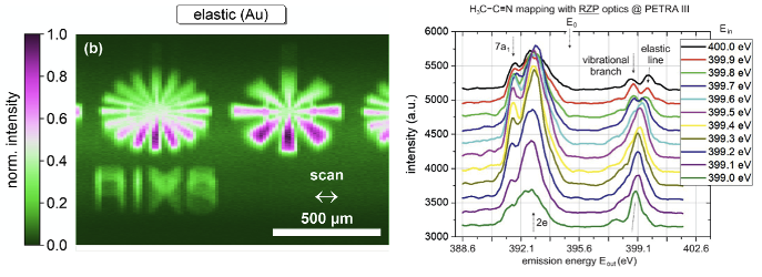
The researchers next replaced the solid test sample with a liquid jet containing acetonitrile dissolved in ethanol. The incident X-ray beam contained a spatially dispersed range of photon energies, forming a “rainbow” along the illuminated section of the liquid jet. The RZP used one detector direction to distinguish the incident photon energies and the perpendicular direction to disperse the emitted X-rays. This allowed the researchers to record a two-dimensional photon-in versus photon-out RIXS map without changing the experimental geometry. The measurement covered emitted photon energies from 388.6 to 401.8 eV and resolved characteristic inelastic, vibrational and elastic features near the nitrogen K-edge. The estimated spectral resolution was ~ 0.3 eV, depending on the incident and emitted photon energies. The full data set was collected in about 220 seconds, much faster than in conventional approaches.
“By combining reflection, focusing, imaging and spectral dispersion in one optical element, the method offers a compact and efficient route to soft X-ray spectromicroscopy and opens new opportunities to investigate matter on the atomic scale,” said last and corresponding author Zhong Yin from SRIS of Tohoku University, who led the international study.
RZPs are manufactured as nm-scale structures on a super-polished Si substrate. Unlike transmission zone plates, they do not require a thin supporting membrane through which the X-rays must pass. Their combination of high dispersion, useful diffraction efficiency and precise manufacturability makes them attractive for photon-intensive soft X-ray experiments. The researchers suggest that the concept could be applied to conventional or coherent X-ray imaging, chemical and elemental mapping, and RIXS spectroscopy. The approach may also be compatible with laboratory high-harmonic sources, laser-driven plasma sources and X-ray free-electron lasers. By combining several optical functions in one component, RZP instruments could reduce the number of optical elements, simplify alignment and enable more time-efficient measurements of spatially heterogeneous materials, liquids and dynamic processes.
- Publication Details:
Title: Experimental verification of soft X-ray imaging and RIXS mapping with a reflection zone plate
Authors: Jens Rehanek, Christoph Braig, Heike Löchel, Felix Marschall, Martin Beye, Dirk Raiser, Sreevidya Thekku Veedu, Benedikt Rösner, Florian Döring, Jens Buck, André Rothkirch, Jens Viefhaus, Christian David, Simone Techert and Zhong Yin
Journal: Optics Express
DOI: 10.1364/OE.596826