Researchers will be able to gain new insights into everything from medical specimens to geological samples, fossils and artworks after a new 4200kg computed tomography (CT) scanner was delivered to the National Imaging Facility node at The University of Western Australia.
The Western Australia National Imaging Facility Node and the Centre for Microscopy, Characterisation and Analysis purchased the large field-of-view Nikon CT (XT H 225 ST) scanner.
Diana Patalwala, from UWA’s Centre for Microscopy, Characterisation and Analysis, said the CT scanner was the first of its type in WA.
“It is the only high-power research dedicated CT scanner in WA, capable of scanning samples as large as 35cm by 35cm by 65cm and up to 50 kilograms in weight,” Ms Patalwala said.
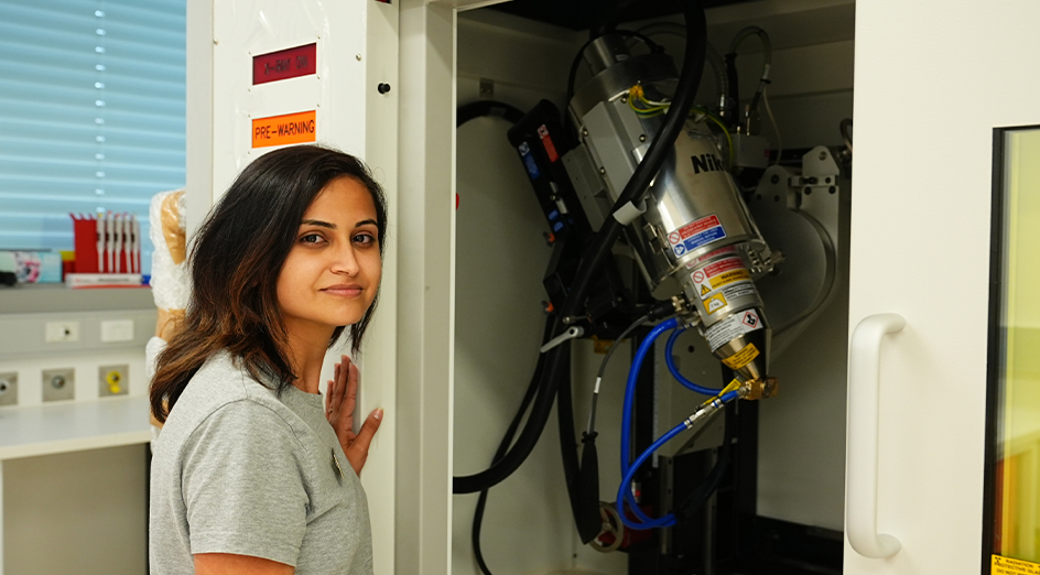 Image: Diana Patalwala, from UWA’s Centre for Microscopy, Characterisation and Analysis, and the new CT scanner.
“The Nikon CT has an extremely high-powered X-Ray source (450W) for penetrating geological, marine and industrial objects as well as the capability of producing lower energy X-Rays (20W), for bio-medical applications.”
The scanner can be used to study inanimate objects such as works of art, statues, pottery and antiques as the penetrating power of the X-rays (225kV/ 450W) can reveal the internal structures of an object without damaging it.
“A CT scan involves X-Rays penetrating an object kept on a rotating stage, with a series of X-Ray images being taken from different angles. Using computer processing it creates 2D cross-sectional images (slices) of the various structures within the object being scanned,” Ms Patalwala said.
CT scanning technique, in combination with advanced image processing software, can also shed light on how an object was manufactured, find evidence of damage and repairs, restoration and even forgery.
“With this CT scanner we are trying to engage with the biomedical, agriculture, environmental, renewable resource, advanced manufacturing, electronics and defence industries,” Ms Patalwala said.
“It will be ideal for scanning archaeological samples, museum specimens and fossils as well. It will get the detailed inside picture without destroying these precious artefacts.”
The scanner, located at Harry Perkins Institute of Medical Research, has been funded by the National Imaging Facility, with the WA State Government and other partner organisations.


